
 Infant Journal
 Browse articles
To 3D or not to 3D? Clinical value of 3D ultrasound in the first trimester
The first commercial 3D ultrasound unit was introduced into clinical practice in 1989.1 Rapid advancements in ultrasound technology, including improvements in machines, software and transducers, have enabled the acquisition of faster and higher resolution 3D images. These advances have facilitated the storage, manipulation and analysis of 3D volumes, providing valuable assistance in accurate assessments and diagnoses of fetal abnormalities. The thermal indices for 3D and 4D ultrasound are comparable to standard 2D ultrasound,2 ensuring safety during its application. The wealth of information stored in 3D volumes allows for the examination of a region of interest in multiple planes,3 enhancing diagnostic capabilities and clinical decision making.
Jayne Lander
Research sonographer, Nuffield Department of Women’s and Reproductive Health, University of Oxford
jayne.lander@wrh.ox.ac.uk
Article reprinted with kind permission from RAD Magazine 2023;49(582):18-19.
Lander J. To 3D or not to 3D? Clinical value of 3D ultrasound in the first trimester. Infant 2024; 20(4): 129-31.
Current practice and clinical value
While 3D ultrasound has the potential for great clinical value, its routine use for sonographers in the UK is not yet widespread. Presently, 3D is commonly employed in specialist fetal medicine scans, but is otherwise often considered to be a novelty to provide keepsakes, often only in private clinics. However, with appropriate training, sonographers can utilise 3D and 4D ultrasound to augment diagnostic capabilities during routine examinations.
Our recent study demonstrated that during the first trimester scan, a volume dataset can be acquired, providing diagnostic views of fetal anatomy in 2D and 3D.4 Eighty-one participants were prospectively recruited and a good quality 3D volume was achievable for 76 of the participants. Diagnostic anatomical views were achievable for the following percentage of cases:4
- axial head (93%)
- facial profile (80%)
- cord insert (80%)
- stomach (62%)
- bladder (22%)
- bilateral hands (92%)
- bilateral feet (80%)
- crown rump length (CRL) (88%)
- nuchal translucency (NT) (72%).
This early acquisition allows for the detection of many anomalies, leading to prompt referral to specialist fetal medicine units. Early diagnosis and information provide expectant parents with more time to make informed decisions regarding their pregnancy.4,5
Furthermore, 3D ultrasound significantly aids in diagnosing or confirming structural abnormalities such as acrania, proboscis, omphalocele, spina bifida, limb deformities and the chorionicity of multiple pregnancies.3
Advanced features such as HDlive (FIGURE 1), silhouette function and skeletal setting allow for impressive image optimisation and the visualisation of fluid-filled structures such as the lateral ventricles (FIGURE 2) or myelomeningocele (FIGURE 3). Spatial temporal image correlation (STIC) can be used to image the fetal heart. By utilising HDlive flow silhouette with STIC there is a good possibility of obtaining five cardiac views at 12-14+6 weeks.6
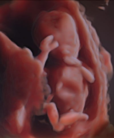
FIGURE 1 3D surface image of a 12-week fetus rendered using HDlive.
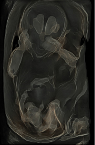
FIGURE 2 Silhouette function demonstrating the lateral ventricles in a 12-week fetus.
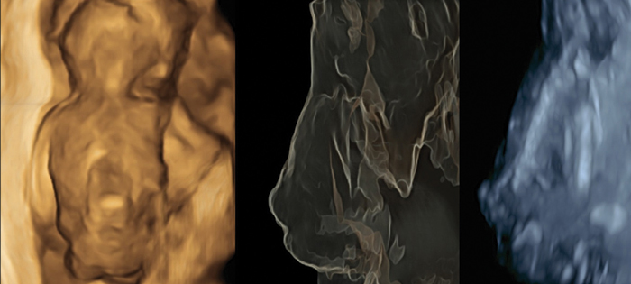
FIGURE 3 Surface, silhouette and skeletal views of a myelomeningocele at 13 weeks’ gestation.
A 3D volume offers a vast amount of information by demonstrating surface anatomy and providing the ability to scroll through individual image slices in sagittal, transverse and coronal planes. The X, Y and Z axes can be adjusted to provide the optimal angle, allowing the acquisition of 2D images of structures in a fraction of the time that it would take to acquire them during a standard 2D scan.
Beyond fetal assessment, 3D ultrasound plays a role in assessment of placental volume and vascularity, and can help to identify placentas that may fail as pregnancy advances. By using power Doppler in conjunction with 3D, the vascular supply of the placenta can be visualised during the first trimester7 (FIGURE 4) and research continues on identifying risk of placental-related complications.
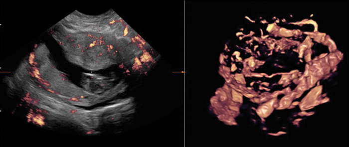
FIGURE 4 2D image on the left and the application of 3D and power Doppler on the right to demonstrate placental vascularity at 12 weeks’ gestation.
Limitations
It is important to highlight that 3D imaging still depends on the angle of insonation, making the acquisition plane (FIGURE 5, sagittal view) of higher resolution than reconstructed transverse and coronal planes. Nevertheless, the resolution of the coronal and transverse planes is often of high enough quality to demonstrate the presence of structures such as the brain, stomach or bladder (FIGURE 6) during a first trimester scan.
In addition 3D volume acquisitions give the ability to measure volumes of the entire fetus or placenta, or individual organs. However, lack of standardisation means that reproducibility of such measurements is generally not adequate for clinical care and efforts to improve this are continuing.8
Fetal movement during acquisition of a 3D volume is generally considered undesirable, but during 4D scanning movements can provide valuable information, in particular when using the cine clip function. Such clips facilitate the assessment of fetal movements and can provide essential information about surface anatomy, presence of limbs, shape of the head and spine and for confirming normal cord insertion in the first trimester (FIGURE 7).
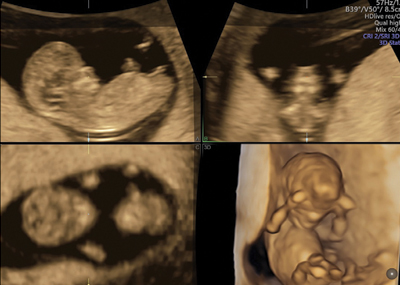
FIGURE 5 Multiplanar view showing sagittal, transverse, coronal and 3D surface images.
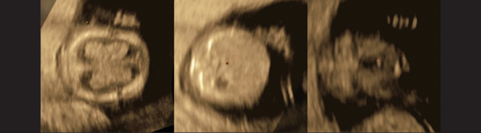
FIGURE 6 2D images of the brain, stomach and bladder extracted from a 3D volume of a 12-week fetus.
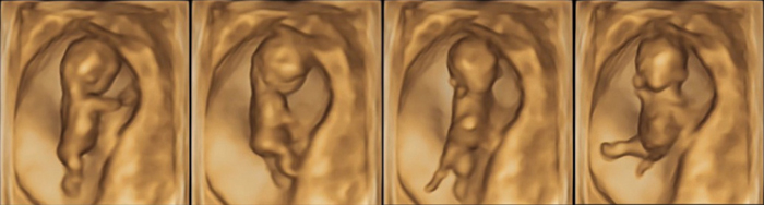
FIGURE 7 Frames taken from a 4D cine-clip which demonstrated both movement and normal surface structures in a 12-week fetus.
Discussion
The clinical value of 3D ultrasound extends not only to specialist scans for identified anomalies but also to routine ultrasound examinations during the first trimester. A skilled operator can optimise the 2D image with an appropriate angle of isonation used to acquire the desired information in the 3D volume. With the fetus still small enough to be contained within one 3D sweep, obtaining multiple desired images, including CRL and NT, is often possible within a single 3D volume in the first trimester.
The examination of surface anatomy and multiplanar 2D slices provides a clearer understanding of the extent of any abnormality, and 3D/4D imaging can be used to confirm suspicions raised during 2D scans. The wealth of information obtained from one 3D volume can be reviewed at a later time, potentially reducing the physical examination time and allowing more time for image review and discussions with parents.
In addition to its diagnostic benefits, 3D ultrasound may help reduce repetitive strain injuries in sonographers. Difficult-to-obtain views with traditional 2D imaging can often be achieved using a 3D volume’s reconstructed image slices, thereby minimising the need for applying pressure and maintaining awkward transducer positions.
Conclusion
The clinical value of 3D ultrasound in obstetrics and gynaecology is substantial and continues to expand with technological advancements. 3D ultrasound provides detailed and comprehensive information that has the potential to enhance diagnostic capabilities and early detection of fetal abnormalities. As with all ultrasound, appropriate training and utilisation of advanced features are needed for image quality optimisation. As the clinical applications of 3D ultrasound continue to grow, it is time to integrate it into routine obstetric practice to improve screening.
 Free download PDF
Or read this article in our
Tablet/iPad edition
- 3D ultrasound technology has rapidly advanced since its introduction in 1989, allowing for faster and higher resolution images.
- 3D ultrasound is not routinely used by sonographers in the UK.
Also published in Infant: