
 Infant Journal
 Browse articles
A case of molybdenum cofactor deficiency type B presenting in early neonatal life
This article highlights an unusual presentation of molybdenum cofactor deficiency in an infant initially presenting with weight loss, possible seizures and feeding. It covers his history, the pathophysiology of the condition and new treatments that have recently become available.
Dr Matthew Rose
Paediatric Specialty Trainee
mrose1@doctors.org.uk
Dr Amy Henderson
Paediatric Neurology Subspecialty Trainee
Dr Nick Wood
Consultant Paediatrician
Dr Chakrapani Vasudevan
Consultant Neonatologist
Bradford Royal Infirmary Neonatal Intensive Care Unit
Introduction
Molybdenum cofactor deficiency (MoCD) is an extremely rare autosomal recessive genetic condition that results in neonatal seizures, severe developmental delay, multicystic leucoencephalopathy and coarse facial features. It has a reported incidence of between one in 100,000 to 200,000.1 There are currently three reported types of MoCD caused by mutations in four known genes (MOCS1, MOCS2, MOCS3 and GPHN), with MoCD Type A (MoCD-A, an MOCS1 mutation) occurring in two-thirds of cases. Molybdenum cofactor is a catalyst for the enzymes sulfite oxidase, xanthine dehydrogenase, aldehyde oxidase and mitochondrial amidoxime reducing component. Deficiency of this cofactor leads to toxic accumulation of neurotoxic and neuroexcitatory metabolites such as sulfite.2 The prognosis of this condition is historically poor, with a median survival of 36 months,3 but recent advances have identified a treatment for MoCD-A that has been shown to improve three year survival rates and symptoms.4 We report on a case of MoCD-B diagnosed in the neonatal period following abnormal brain imaging.
Case report
This case follows a male term baby, born at 39+5 weeks’ gestation by spontaneous vaginal delivery, after a low risk pregnancy. There were no risk factors for infection. Of note, there was a family history of consanguinity and a previous sibling had died of unknown causes around eight months of life. He initially presented to neonatal services at three hours of life, with tachypnoea, hypoxia and possible seizure activity (initially reported as hypertonic, and at time of neonatal review felt to be hypotonic). He was admitted to the neonatal unit for observation. Initial blood tests showed normal electrolytes and a lactate of 2.5. There was no further seizure activity observed, and he was discharged after establishing feeding. Because of the initial concerns regarding tone and possible seizures, an outpatient cranial ultrasound scan and secondary care follow-up were arranged.
On day five of life, he presented to the general paediatrics ward after a referral from community midwifery who were concerned about jitteriness. He was examined and felt to be clinically well, had a normal blood gas, and was discharged home. He was referred to paediatrics again on day nine of life with 11.5% weight loss. He was again assessed and felt to be well. Routine biochemistry bloods were sent, which were unremarkable. He was discharged home. He presented for a third time to paediatrics on day 12 of life, this time with a total weight loss of 14.6% since birth. Routine biochemistry and a blood gas were again normal, and he was discharged home with a feeding plan. He did not present to paediatrics again before his imaging was performed.
A cranial ultrasound was performed as an outpatient on day 20 of life. It was grossly abnormal, showing a possibly absent corpus callosum and bilateral abnormalities in both frontal-parietal regions, possibly cystic. An MRI was urgently arranged and performed three days later. This showed a thinned corpus callosum, extensive cystic abnormalities throughout both hemispheres, and abnormal signal in the basal ganglia (FIGURE 1).
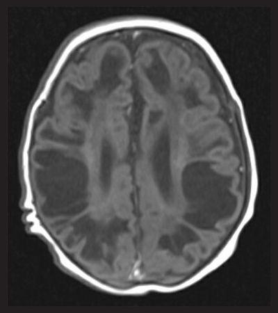
FIGURE 1 Axial T1 MR image showing extensive cystic changes throughout the brain parenchyma.
Further investigations were sent including routine biochemistry bloods, a metabolic screen (lactate, ammonia, plasma amino acids, urine organic acids, acylcarnitines and very large chain fatty acids) and genetics (array comparative genomic hybridisation (CGH) array and DNA for storage). Routine biochemistry was again normal. His ammonia was normal (66). His CGH array was normal. His plasma amino acids profile showed a low cysteine (<2, reference range 21-73). The clinical biochemistry team noted this and added a urate level to aid inter-pretation. His urate level was undetectable (<30, reference range 200-430). These results were felt to be consistent with molybdenum cofactor deficiency. He was referred to the regional metabolic team and after a short inpatient stay with a failed trial of treatment, was diagnosed with MoCD-B. He was discharged home under the care of community paediatrics and the specialist metabolic team for further follow-up.
Discussion
Our case is worth noting as the presentation does not fit with the most commonly reported symptoms in MoCD. These are neonatal refractory seizures and neonatal encephalopathy, sometimes mimicking hypoxic ischaemic encephalopathy.1,5 In our case, however, failure to thrive secondary to feeding difficulty was the main symptom. There was no reported seizure activity apart from possibly immediately at birth, and there were never concerns regarding encephalopathy. There was also no mention of coarse facial features, another commonly reported finding. Previous case studies have reported that seizures occur in all cases of MoCD, whereas feeding difficulty is reported in around two thirds of cases.6
A new treatment for MoCD-A has recently become available. In MoCD-A, the MOCS1 gene mutation stops the conversion of guanosine triphosphate to cyclic pyranopterin monophosphate (cPMP), which itself is needed to produce molybdenum cofactor. Fosdenopterin is a synthetic form of cPMP that is administered as a once daily infusion. It has been shown in an early clinical trial to increase life expectancy in patients with MoCD-A. In regulatory trials, Fosdenopterin-treated patients had a three year survival probability of 84%, compared to untreated patients who had a three year survival probability of 55%.7 Unfortunately, it has no effect on MoCD-B or MoCD-C, as the mutations causing these diseases affected the molybdenum cofactor synthesis pathway further downstream, after cPMP has already been produced (FIGURE 2).
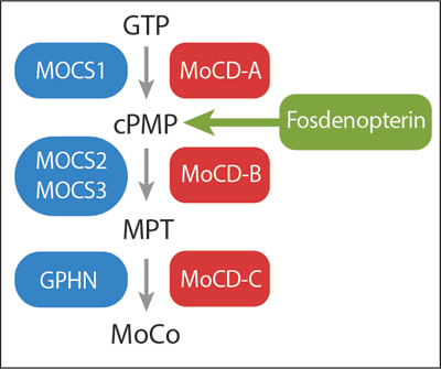
FIGURE 2 The synthesis of MoCo, the enzymes needed for each step (blue boxes), the disease caused by loss of enzyme function (red boxes), and the step in the synthesis pathway where Fosdenopterin has its action. GTP = guanosine triphosphate, cPMP = cyclic pyranopterin monophosphate, MPT = molybdopterin, MoCo = molybdenum cofactor.
Conclusion/learning points
While the diagnosis of MoCD is ultimately confirmed with genetic testing, it is worth noting that there is a classical biochemical profile that can be identified in those affected. Due to the loss of function in the enzyme xanthine dehydrogenase, shortly after birth, patients with MoCD will have nearly undetectable levels of uric acid in their serum. They will also have raised urinary xanthine and hypoxanthine levels. In our case, it was the biochemistry team that noted the low cysteine levels, which prompted them to test for uric acid. Low cysteine levels are due to accumulated sulfite linking with cysteine to form s-sulfocysteine, an alternative diagnostic marker.8
Infants presenting with weight loss early in life are a common presentation to paediatric wards. A structured assessment to these patients is crucial. While issues such as mechanical feeding difficulty and breast milk supply problems are common, pathological causes need to be considered also. There are a multitude of diagnoses that could lead to faltering growth. In cases such as this child, it could have been worth considering second line investigations such as metabolic screening around his third presentation with persistent weight loss.
While MoCD is extremely rare, the emergence of new therapies that can potentially extend a patient’s life highlights the need to be aware of it as a possible cause of seizures, encephalopathy and weight loss in the newborn infant.
 Free download PDF
Or read this article in our
Tablet/iPad edition
- Molybdenum cofactor deficiency is associated with seizures, encephalopathy, coarse facial features, developmental delay and feeding difficulties.
- There is a new treatment available for molybdenum cofactor deficiency type A that can improve three year survival in patients.
Also published in Infant: