
 Infant Journal
 Browse articles
Back-to-sleep or tummy time? Positioning of premature infants and its impact
The primary goal of infant positioning in the neonatal unit is to mimic the in utero environment. This article will consider this in relation to musculoskeletal development, cerebral perfusion, sleep promotion and cardiorespiratory stability. The practical application of positioning ideals will also be addressed.
Fiona Allison
Staff Nurse
Surabhi Kumar
Paediatric ST1
Raju Sunderesan
Consultant Neonatologist
Ayrshire Maternity Unit, University Hospital Crosshouse, Kilmarnock, Scotland
The positioning of the premature infant in the neonatal unit is a vital consideration in reducing morbidity and mortality. Position and handling forms a vital part of the Neonatal Integrative Developmental Care Model, and it impacts all areas of neonatal development. The use of different positions in infants has been postulated to improve outcomes in other areas of neonatal development.
This paper explores studies and reviews regarding infant positioning and its effects on cardiorespiratory stability, musculoskeletal development, sleep and cerebral blood flow. Literature searches were carried out using PubMed, MEDLINE and Cochrane database searches to find both clinical studies and systematic reviews.
Study review
A number of studies and meta-analyses have found that the prone position improves cardiovascular stability, especially in ventilated infants. This position has also been shown to improve sleep, more so when used in conjunction with nesting and swaddling. Smaller trials indicate that the stability of cerebral blood flow may be improved with infants nursed in the prone and/or inclined position, leading to decreased rates and severity of peri/intraventricular haemorrhages. Development of neuromuscular pathways and musculoskeletal stability in neonates is significantly improved by the employment of the flexed, midline position, with the use of barriers that mimic the uterine wall. These are further improved by the implementation of physical activity programmes. The implementation of nursing care bundles that encourage frequent position changes and handling lead to significant improvements in neonatal outcomes.
Vital considerations in positioning are creating a flexed, midline position in neutral alignment, the use of boundaries and, most importantly, maximising comfort for the infant. The emphasis on these criteria as part of a nursing care bundle improves neonatal outcomes and has positive impacts on the infant’s cardiorespiratory stability, musculoskeletal development and neurovascular development.
In utero, the fetus has its own tailor-made incubator. It is protected from all external stimuli such as physical sensations, bright lights and loud noises. Its skin, muscles and bones are protected from external forces as it floats in its bubble of amniotic fluid. When babies are born prematurely, neonatal teams strive to provide an environment as close to the in utero milieu as possible. Medical advancements have enabled survival of infants at extreme prematurity. The youngest was born at just 21+1 weeks’ gestational age (GA).1 This is not, however, without the risk of long-term poor neurodevelopmental outcomes.2
The positioning of a neonate in the incubator is a vital consideration in reducing morbidity and mortality. Premature infants have immature self-regulatory behaviour and lack the ability to self-soothe when exposed to stressful external stimuli.3 Prone positioning is often employed in neonatal units and has been found to improve oxygenation and decrease respiratory distress.4 Prone positioning and the use of nests as postural support has been shown to improve sleep and reduce distress behaviours exhibited by these infants.5
This review of current evidence for neonatal positioning practices explores the impact of position on the infant’s cardiorespiratory stability, musculoskeletal development, sleep and cerebral blood flow. These aspects, in combination, give evidence on the optimum strategies for developmental positioning and the basis for future neonatal care guidelines.
Principles of neonatal care
The synactive theory was developed by Heidelise Als, an American neuropsychologist based in Boston, who observed preterm behaviour.6 The theory provides a framework to conceptualise the neurobehavioural capabilities of the fetus and newborn. This theory describes the infant in four domains of functioning that develop throughout gestation, and their interaction with the environment – from within the womb to the ex utero neonatal intensive care unit (NICU) (FIGURE 1).
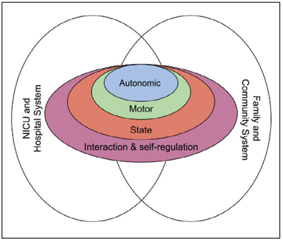
FIGURE 1 The synactive theory of individualised neonatal care, adapted from Als.10
Als and McAnulty7 used the synactive theory to develop NIDCAP (The Newborn Individualized Developmental Care and Assessment Program). This theory was further built on by Altimier and Phillips8 who created the Neonatal Integrative Developmental Care Model. This stipulates seven core measures for infant-centred care, further expanding on the external factors affecting the neonatal journey (FIGURE 2).
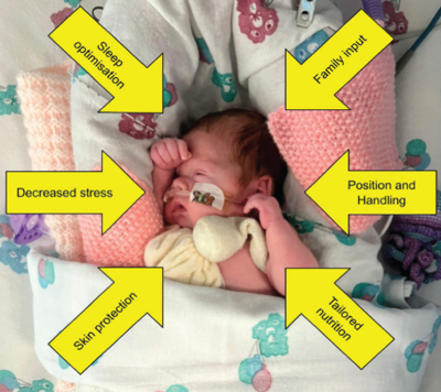
FIGURE 2 Neonatal Integrative Developmental Care Model, adapted from Altimier and Phillips.
As detailed above, there are numerous factors that must be taken into account in care of the neonate. While the Neonatal Integrative Developmental Care Model considers positioning as its own core measure, it uniquely affects each of the other measures. Optimisation of position improves sleep and decreases stress, can be tailored to protect skin and requires family input, thereby substantiating the parental role as the primary carers.
Positioning and cardiorespiratory stability
Satisfactory cardiorespiratory function requires mature lungs capable of gas exchange. A successful transition from fetal circulation is necessary and the underlying homeostatic and neurological mechanisms to support these physiological processes need to be working. Premature birth denies these infants their optimal physiological state and consequently they suffer varying degrees of cardiorespiratory instability. Positioning, in conjunction with supportive medical interventions, can lead to improved stability in cardiac and respiratory function.
A number of studies have shown that nursing infants in the prone position leads to improvements in an infant’s cardiac and respiratory status. A crossover study of 40 ventilated infants by Hassan et al9 demonstrated that prone positioning led to a significant improvement in oxygen saturation, with stable heart rate and breathing, when compared with the supine position. Rather than measuring clinical parameters, Dimitrio et al10 assessed work of breathing by monitoring diaphragmatic activity in 40 preterm infants breathing room air (28-34 week GA). They compared prone, flat-supine and inclined-supine (45 degree tilt) positioning and concluded that energy expenditure by the diaphragm was lower while prone and inclined-supine, when compared to flat supine. This effect was more evident in previously ventilated infants.
Prone positioning appears to decrease the degree of thoraco-abdominal asymmetry and asynchrony due to lowered chest wall compliance, which then allows for increased tidal volumes. The anterior chest wall expansion is restricted, which results in a more homogenous chest wall compliance, enabling greater recruitment of the posterior zones. Prone positioning reduces the compressive force from the abdomen, thereby improving inferior movement of the diaphragm. This increase in tidal volume also decreases the impact of gastric distension by increasing downward diaphragmatic compression (FIGURE 3).
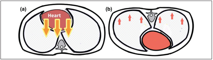
FIGURE 3 Physiological dynamics in (a) Supine position – there is gravitational pressure of the heart on the lung, and there is less homogenous compliance of the chest wall. (b) Prone position – the gravitational pressure of the heart is reduced. There is more homogenous compliance of the chest wall due to the restriction of anterior chest wall movement.
The inclined position was further investigated by Li et al,11 who found that higher levels of oxygenation were associated with positioning on a 15 degree tilt in both prone and supine, but that comparatively, the former had a greater positive effect, with an additional reduction in respiratory rate and blood pressure. This is hypothesised to be a result of improved ventilation of lower lung segments, a more optimal pulmonary ventilation to blood flow ratio, and decreased intra-abdominal pressure when in the elevated position.
Improved pulmonary function was also demonstrated in a study by Gouna et al.12 In this crossover study, infants receiving nasal continuous positive airway pressure (nCPAP) were found to have higher tidal volumes and oxygen saturations, with fewer apnoeic events, when nursed prone or in the left lateral position (as compared to supine). There were, however, no differences noted in respiratory rate, heart rate or blood pressure between the three positions.
Implementation of the prone position for infants with respiratory distress is common practice. However, the use of inclined or lateral positions is less commonly seen. A 2016 Cochrane review by Rivas-Fernandez et al13 concluded that prone positioning leads to significant improvement in outcomes of infants receiving mechanical ventilation. They did not, however, find any significant benefits to lateral or elevated positioning, indicating this is an area requiring further research.
Musculoskeletal development
Fetal movements commence at about seven weeks of gestation. With advancing gestation, however, freedom to move becomes increasingly restricted, due to increasing fetal size and decreasing amniotic fluid. In the third trimester, this restrictive fluid-filled uterus supports the fetus in a flexed midline resting position. The initial dominance of flexion allows the infant, once exposed to gravity and free movement, to gradually develop balanced anti-gravity movements.14 Movement against the recoiling uterine wall also promotes bone mineralisation15 and the moulding of joints,16 while the flexed midline position promotes self-regulatory behaviours such as hand to mouth, which helps reduce fetal stress.5
Goniometric assessment of premature infants has shown that flexor tone and recoil of the lower extremities develops about 33 to 35 weeks, while in the upper limbs it is about 35 to 37 weeks.17 When born prior to this process, the infants’ pattern of maturation can be both positively and negatively influenced by the ex utero environment. Preterm birth results in a reduction in bone loading that is usually afforded by the uterine wall.15 When coupled with the reduced efficacy of ex utero calcium absorption, this leads to the development of osteopenia.
The premature infant’s poor tone, in combination with the above, forces the infant into extended resting postures. This in turn alters the direction of the neurological processes that underpin muscular control, with the neural pathways supporting extensor positioning becoming relatively more dominant.14 Ortqvist et al18 demonstrated that by three months of age, premature infants were more likely to show atypically extended limbs, head asymmetry and had poorer motor fluidity than their term counterparts.
According to Sweeney and Gutierrez,16 suboptimal posturing of the preterm infant can negatively impact their longer-term development. Hyperextension of the neck, with associated shoulder retraction, can impede later acquisition of head control, downward gaze and hand to midline movements. It also reduces upper limb stability when prone, which may delay the ability to roll over. Infants left in the ‘frog-leg’ position (abducted hips with externally rotated feet) have been demonstrated to have difficulties with crawling and position transitions, as well as gait abnormalities. Samsom and deGroot19 studied 75 infants (less than 33 weeks’ GA) and also found that poor posturing in the neonatal period was predictive of suboptimal gross and fine motor abilities at 12 months corrected age (independent of the incidence of prematurity-related brain injury).
Use of aids to minimise cranial moulding have been shown to have some efficacy in reducing incidence of plagiocephaly. Degrazia et al20 compared the use of mouldable pillows with or without the addition of a specifically designed ‘cranial cup’ in 62 infants. They concluded that the combination of the two aids reduced the incidence of cranial deformity in infants corrected to term. A further study by Uchio et al,21 which continued the use of a positioning pillow until six months corrected age, reported a reduction in the development of plagiocephaly.
Research of positioning interventions for the promotion of musculoskeletal development has demonstrated some positive outcomes. Stalnaker and Poskey15 analysed the findings of 12 trials that evaluated the effect of physical activity programmes for infants between 26-35 weeks’ GA. These programmes lasted up to eight weeks and demonstrated an improvement in bone density. Similarly, Hughes et al22 undertook a meta-analysis of trials, totalling 6,234 subjects, comparing the efficacy of targeted motor interventions against general developmental care practices. They found that targeted interventions led to a significant improvement in motor function, including improvement in the age of reaching motor milestones, such as walking. This effect appears to be most evident at three months’ chronological age, but continues throughout the first two years of life.
The interventions that correlate best with improved motor outcomes are those that promote hand to midline orientation and those that enable regular movement opportunities in the supine, prone and lateral positions.14,15,22 These findings indicate the need for a structured approach to premature infant positioning in the NICU.
Sleep
Functional and structural maturation of the fetal brain occurs primarily during sleep states. In the second trimester, neural network establishment occurs with the development of thalamocortical and cortico-cortical connections. This is coupled with the pruning of connections based on initial endogenous and subsequent exogenous neurosensory activity.23
These processes occur in a period of indeterminate sleep. Further synaptogenesis, however, requires the maturation of sleep into distinct rapid eye movement (REM), or non-rapid eye movement (NREM) cycles, which begin from 28 weeks’ GA. During REM sleep, endogenous stimuli arising from the rudimentary neurosensory system instigate further synaptogenesis. This prepares the brain for ongoing development through exposure to exogenous stimuli, such as maternal voice or touch. NREM sleep performs an equally important role. In conjunction with REM sleep, NREM is central to the consolidation of memory, learning and the promotion of neural plasticity.24 Term infants have been found to spend their sleep equally between REM and NREM, while in adults around 80% of sleep is NREM. Conversely, fetuses from 28 weeks’ GA show a predominance of REM sleep, in order to accommodate the need for rapid brain development in the third trimester, as depicted in FIGURE 4.25
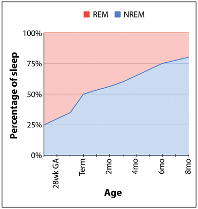
FIGURE 4 Sleep pattern with age.
Premature infants usually spend up to 90% of their time asleep to optimise brain development.26 Functional MRI scans of premature infants’ brains show differences in functional brain connectivity as compared to term infants. This is particularly evident in sensory and stress processing regions, such as the hippocampus and insula, thought to be a result of the dramatically increased exposure to stressors in the ex utero environment. Placental steroids, shown to aid white matter maturation and modulate fetal sleep, are lost in premature infants. This change in sleep modulation and the less-than-optimal ex utero environment impacts on further synaptogenesis and thus a cycle of perpetuating neurological compromise ensues.26
One of the strategies used to promote this essential sleep and to reduce environmental stimuli is the use of developmental positioning. Peng et al27 studied how positioning influences the infant’s sleep state when exposed to environmental stressors. They concluded that prone positioning, as compared to supine, elicited fewer stress responses, which consequently enabled longer periods in quiet sleep.
Similarly, Kihara and Nakamura28 demonstrated longer periods of quiet sleep among premature infants nursed prone with positioning supports, compared with those in the prone position without support. This effect was thought to be due to enhanced physiological flexion in the supported prone position. Other positioning strategies such as swaddling and nesting have also been demonstrated to induce longer periods of quiet sleep.
Cerebral blood flow
Survival of extremely preterm infants (born at less than 28 weeks’ GA) has increased sharply in the last decade, which has been accompanied by higher rates of subsequent neurodevelopmental disability. The most notable pathological correlate for neuro-morbidity includes periventricular leukomalacia and germinal matrix periventricular and intraventricular haemorrhage (PIVH). The preterm brain is more susceptible to these injuries, because of both hypoxia and dysregulation of cerebral perfusion.29
Cerebral blood flow (CBF) relies on cerebrovascular pressure autoregulation in order to remain unchanged during fluctuations in blood pressure. In 1959, Neils Lassen first illustrated the cerebral autoregulation curve in adults with optimal cerebral blood flow occurring within a cerebral pressure range of 50 to 150mmHg.30
On the contrary, the autoregulatory curve in term neonates is maintained within a narrow arterial pressure range of 20 to 60mmHg.31 Beyond these limits, CBF becomes pressure passive, resulting in steep changes with a linear correlation between CBF and systemic blood pressure. In preterm infants, this plateau range is narrower and lower still at around 20 to 40mmHg and varies significantly. Using transcranial Doppler to assess CBF, Rhee et al32 found that autoregulatory function is poorer at lower gestational age. These factors lead to an increased risk of both hypo and hyper-perfusion injuries, most notably PIVH.33 This is depicted in FIGURE 5.
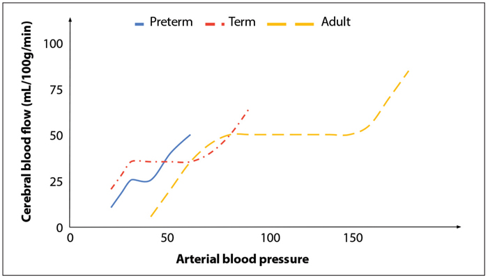
FIGURE 5 Cerebral blood flow changes with arterial blood pressure. Comparison between premature infants, term infants and adults is depicted.
A randomised controlled trial by Kochan et al40 compared head elevation of 30 degrees with supine position in 180 extremely low birth weight infants in the first four days of life. Although there was no significant difference in the incidence of all PIVH between groups, those with elevated head positions developed significantly fewer grade 4 PIVHs.
Spengler et al34 carried out a smaller study of 21 stable infants (26-32 weeks’ GA) using near-infrared spectroscopy to measure CBF at cerebral tissue oxygenation. They compared supine positioning with the head in the midline and prone positioning with the head turned 90 degrees and found no significant change in CBF or cerebral tissue oxygenation.
A 2020 Cochrane meta-analysis of trials found no significant evidence that a change in head position reduces mortality, or the incidence of PIVH.35 A similar conclusion was reached by a more recent systematic review by Jani et al.36 This review did, however, conclude that while there were no statistically significant positive findings, there was little to no evidence of any negative effects on the prone or lateral position, when compared to supine.
Developmental positioning and the use of aids
As discussed above, there are several ways in which the positioning of a premature infant impacts their physical and physiological state. Developmental positioning refers to the use of positioning as a tool to optimise care and aims to mimic the protective and adaptive characteristics of the uterus.8 Appropriate flexed positioning, protection of sleep and minimisation of cerebral blood flow fluctuations can, to some degree, mediate the potentially neurotoxicex utero environment.
An ‘ideal’ positioning aid, therefore, should promote proprioception by providing a close circumferential high boundary to allow for recoil of limbs on extension and support the infant in resting physiological flexion (FIGURE 6).
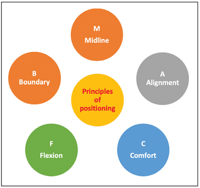
FIGURE 6 Principles of ideal neonatal positioning.
There are a variety of aids utilised across neonatal units. Homemade ‘nests’ provide an outer boundary. These are rigid and of a standard shape and size, which limits their functionality. There are, however, various commercial aids that allow for a greater degree of customisation. These include size-specific nests with adjustable straps, mouldable pellet-filled boundaries and fluidised positioners, which have the added benefit of keeping their shape against gravity. Additional support can be added by using ‘insert’ positioners. A prone positioner, for example, allows the infant to maintain a flexed position and prevents pressure on the knees. Similarly, a beanbag insert can be used to provide more contoured support to a laterally positioned infant or can be used to support the head in midline. These can also be used to provide gentle weighted containment that mimics the containment hold routinely used by caregivers and which has the same calming effect as swaddling. There are also commercially produced structured swaddling aids, which promote recoil of the limbs back into physiological flexion.
Research has demonstrated that targeted education and the use of positioning tools in the neonatal unit are associated with improved and more consistent practice among staff.37 The Infant Positioning Assessment Tool (IPAT) by Koninklijke Philips and its numerous adaptations have been used in many quality improvement initiatives (FIGURE 7).38 The IPAT’s pictorial table provides an excellent guide on the ideal head-to-toe positioning, with a corresponding scoring system that encourages staff to assess and optimise their own practice and has been shown to improve neonatal outcomes.38,39
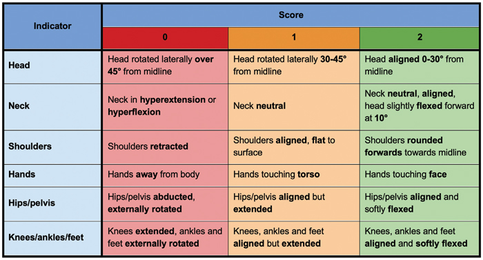
FIGURE 7 Infant Positioning Assessment Tool. A score between 0-2 is given for each body part, and a total score calculated. A score of 12 is ideal; 9-11 is acceptable, ≤8 indicates a need for repositioning. Adapted from Coughlin et al.38
The ideal positioning aid should provide a close circumferential boundary, with high enough walls to allow for recoil of limbs on extension (thereby promoting proprioception for neurological development) and should be able to support the infant in resting physiological flexion while supine, prone and lateral. From a practical perspective, any positioning aids must also be able to accommodate medical devices and allow for adequate observation and access to the infant. Ideally the caregiver should have access to a variety of aids in order to accommodate each infant’s individual developmental and clinical needs.
It is worth noting that the introduction of aids and positioning tools, while essential for changing practice, should first be preceded by a staff understanding of the physiological and clinical impacts of positioning. This knowledge should provide incentive, as well as promote confidence, for staff having to change their established practices. Indeed, one of the barriers to change recognised in quality improvement projects was staff reservations surrounding the need for change in the first place.37 The actual practicalities of introducing or changing care interventions will also impact on the likelihood of success. The time demands of existing unit activities makes dissemination and consolidation of new learning, as well as training in new equipment, difficult to accommodate. Cost implications of introducing new products and the practical issues of storage, maintenance and compatibility with existing equipment must also be given due consideration. In the presence of financial restraints, having a theoretical framework on which to base positioning practices should support staff in utilising existing resources, such as individualising homemade nests or using rolled up linens as positioning inserts. Visual aids such as posters of good positioning can provide simple advice to members of the team carrying out position changes (FIGURE 8).
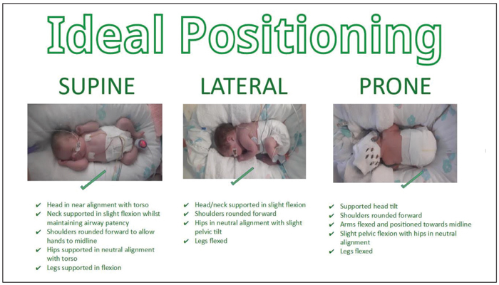
FIGURE 8 ‘Ideal positioning’ poster describing optimal supine, lateral and prone positioning.
Conclusion
Evidence shows that the well positioned preterm infant enjoys longer periods of quiet sleep, thus optimising brain development, and experiences less cardiorespiratory instability, conserving energy for growth. Some research indicates that containment, similar to that experienced in utero, promotes musculoskeletal function. There is limited evidence, however, supporting a causal connection between positioning and fluctuations in cerebral blood flow.
The practical application of developmental positioning can be promoted using positioning aids and qualitative tools. Their use must be supported by an understanding of the physiological impact in order for optimal benefit to be obtained.
What does remain a contentious issue, however, is the extent to which developmental care practices impact on long-term developmental outcomes. Several reviews have concluded that these effects may be relatively small and limited to the first two years of life.40
It could be argued that the many confounding variables that impact on long-term outcomes make it impossible to ascertain a simple cause and effect for such interventions. Furthermore, the fact that interventions such as position optimisation do have demonstrable immediate physiological and emotional effect on the hospitalised infant supports its continued use as a point-of-care intervention within the NICU.
 Free download PDF
Or read this article in our
Tablet/iPad edition
- Fetal positioning in the womb enables optimal neurological and physical maturation. Ex utero positioning of premature infants should aim to mimic this environment.
- Ex utero positioning can influence the short and longer-term developmental outcomes of preterm infants.
- Optimal positioning requires an understanding of normal in utero neurodevelopment and recognition of how this may be compromised by premature birth.
- Application of positioning principles should be supported with guidelines and positioning tools.
Also published in Infant: