
 Infant Journal
 Browse articles
Isolated radial nerve injury in newborns: Highlights on diagnosis
Isolated radial nerve palsy is rare and must be differentiated from obstetric brachial plexus palsy to ensure the correct course of action. The authors present a case of isolated radial nerve palsy in a female neonate, with subsequent treatment and recovery, and a literature review.
Moustafa Eldalal
Paediatric Senior Clinical Fellow
moustafa_badreldin@outlook.com
or moustafa.eldalal@nhs.net
Michael Gunn
Paediatric Junior Clinical Fellow
Siddhartha Paliwal
Paediatric Consultant and Neonatal Lead
Queen Elizabeth Hospital, Lewisham and Greenwich NHS Trust
Eldalal M., Gunn M., Paliwal S. Isolated radial nerve injury in newborns: Highlights on diagnosis. Infant 2024; 20(4): 116-19.
Background
Neonatal nerve injuries of the upper limb may have different causes. The most common cause is obstetric brachial plexus palsy (OBPP) in which Erb’s palsy is more common than Klumpke’s palsy. However, radial nerve palsy is very rare with only a small number of cases reported. Hence, it is likely to be misdiagnosed as OBPP. In carrying out our literature review, we have found 84 cases previously reported.
As radial nerve palsy is uncommon, paediatricians need to be aware of the condition in order to identify it, to provide appropriate management and to plan the necessary follow-up appointments. Although radial nerve palsy has varying causes, isolated radial nerve palsy is most commonly associated with a difficult delivery1 and has an excellent prognosis. Therefore, accurate diagnosis will enable paediatricians to reassure parents and provide an accurate prognosis.
Case presentation
We present a case of isolated radial nerve palsy in a female neonate, born to a primigravida mother at term (40 weeks of gestation) weighing 3.7kg at birth. She was born by forceps delivery due to failure to progress to the second stage of labour. The baby required drying, stimulation and one set of inflation breaths at birth. Her Apgar score was 6, 9 and 10 at one, five and 10 minutes respectively.
On routine NIPE examination (newborn and infant physical examination) the following day, the baby had left wrist and finger drop (FIGURE 1). She had a full active range of movement of the right upper limb at all joints. She also had a full active range of movement on the left shoulder and arm against gravity. There was marked left wrist drop and finger drop with no active movement of wrist extension, even with stimulation. Furthermore, there was no active finger extension, but a minimal flicker of finger extension on stimulation. Passive movement of left wrist and fingers were normal with full range of movement. The baby had Moro reflex and palmar grasp reflex bilaterally. Therefore, it was clear that it was a distal injury not involving the shoulder or arm of the left upper limb.
On inspection of the left arm, there was an area of redness at posterolateral aspect. It was a firm non-pitting nodule in the subcutaneous tissue, which is consistent with fat necrosis (FIGURE 2).
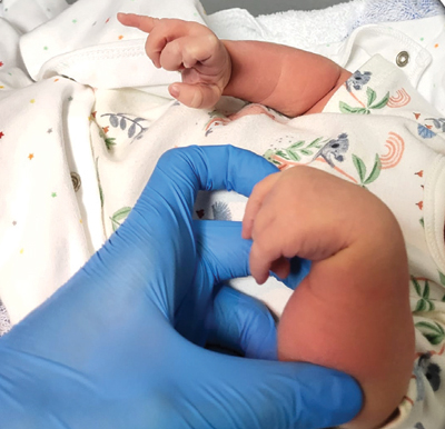
FIGURE 1 Left wrist and finger drop with inability to extend wrist, metacarpophalangeal or interphalangeal joints. Normal wrist and fingers extension of the right hand
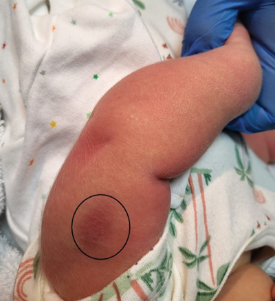
FIGURE 2 Red area at the posterolateral aspect of the left arm, indicative of fat necrosis.
Investigations
The baby had x-rays to exclude humerus fracture, which came back normal (FIGURE 3). At six weeks of age, nerve conduction studies and electromyography suggested mild radial nerve affection with good recovery. This was expected due to the time lag between when the injury was sustained and when the investigations were carried out.
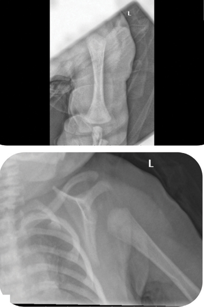
FIGURE 3 Normal x-ray of the humerus.
Differential diagnosis
Other pathologies can be associated with radial nerve injury in neonates such as humerus fracture5 and infantile cortical hyperostosis,20 which can be easily excluded by an x-ray of the arm. Other causes that are described in the literature include angioleiomyoma,21,22 amniotic constriction band,23 intrauterine stab injury,24 septic shoulder25 and use of the blood pressure cuff in premature infants.26
The management in these cases is dependent upon the cause. In some case, surgical intervention may be necessary.
Treatment
Before discharge from the postnatal ward, the baby was referred for physiotherapy. Her parents were taught to perform passive stretches (wrist extension, finger extension, finger abduction and thumb abduction). They were encouraged to perform these exercises regularly, to coincide with nappy changes. Sensory stimulation was also demonstrated and a follow-up up appointment was arranged.
Outcome and follow-up
At two months of age, the baby presented for her follow-up appointment. There was no wrist or finger drop. Her left wrist and digital extension were almost comparable to that on the right side. There was also a significant improvement in left finger power. However, it was still slightly weaker than the right side. Therefore, the decision was made to continue physiotherapy until full recovery of strength on the left side was achieved. This was achieved by six months of age and she was discharged from physiotherapy.
Discussion
Epidemiology
Isolated radial nerve palsy is rare in newborns. We carried out a literature search for the number of cases reported in literature using a PubMed search of “radial nerve and newborn”; “radial nerve and neonate”; “wrist drop and neonate” and “wrist drop and newborn”. We found 84 cases reported before our case, excluding cases where there was other pathology causing radial nerve injury.20-26
Pathophysiology
The pathophysiology of isolated radial nerve palsy is thought to be caused by compression of the distal part of the arm at the location of the spiral groove. This is where the course of the radial nerve is most superficial.
There is data to suggest that the compression can occur either before the onset of labour while the baby is intrauterine, or during labour itself. In one case study, neurophysiology studies showed a denervation pattern suggestive of the injury occurring before onset of labour.3 Hence, it was suggested that it was caused by prolonged compression of the nerve at the lower part of the arm against the pelvic brim, either in utero or during labour.3 This reasoning is supported by the presence of a skin lesion at the postero-lateral aspect of the arm where the radial nerve passes in 71.7% of cases reported in the literature. This skin lesion can be a bruise, ecchymosis or more commonly, fat necrosis.4 These are thought to be consequences of the prolonged compression that causes the skin lesions as well as the radial nerve palsy.
Clinical picture
Isolated radial nerve palsy presents with wrist and finger drop with inability to extend the wrist, metacarpophalangeal joint and interphalangeal joints. However, normal full range of active movement will be present in both the shoulder and the arm. Palmar grasp reflex will also be present.2
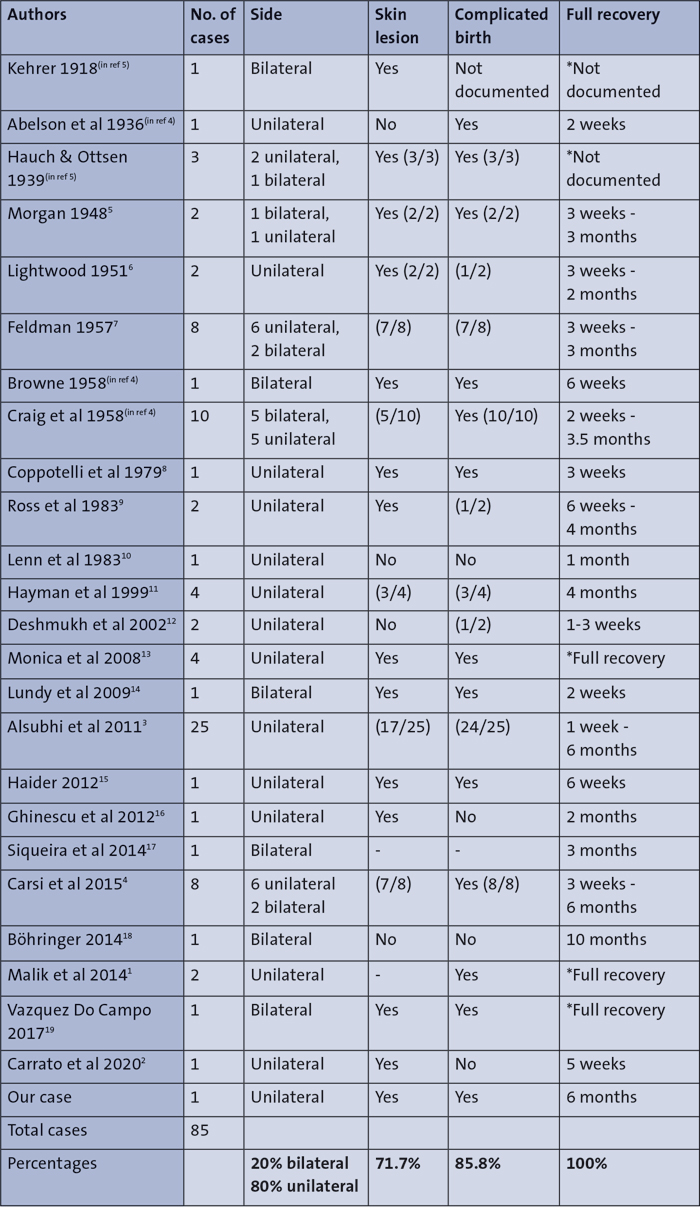
TABLE 1 Cases of isolated radial nerve palsy described in the literature. (*) No clear documentation on when full recovery occurred. (-) No documentation about presence or absence of skin lesion/circumstances of birth.

TABLE 2 Simplifying differences between causes of OBPP and isolated radial nerve palsy.
Review of isolated radial nerve injury in the literature
Assessment and diagnosis
Differentiating obstetric brachial plexus palsy from isolated radial nerve injury is important because it will help in explaining the prognosis to the parents and providing reassurance. Erb’s palsy presents with loss of shoulder abduction, loss of shoulder external rotation and loss of arm flexion, which is described as “waiter’s tip wrist”.27 These babies will have abnormal Moro reflex which should raise the suspicion of Erb’s palsy. However, the exclusion of clavicular fracture is impor-tant as it can cause pseudoparalysis, which can be easily mistaken for Erb’s palsy.27
Klumpke’s palsy affects the muscles of the forearm and hands. Hence, it presents with wrist extension, extension at metacarpophalangeal joints and flexion at interphalangeal joints, which gives the appearance of a “claw hand”.28 It is worth mentioning that it may be associated with Horner syndrome (ptosis, miosis and anhidrosis) as the injury might involve T1 nerve root.28 Palmar grasp reflex is absent in these babies, as ulnar and median nerves are involved in the reflex29 and both will be injured in Klumpke’s palsy.
Prognosis
Isolated radial nerve palsy is rare. However, its prognosis is excellent with complete recovery by six months without needing surgical intervention. In contrast, other causes of radial nerve injury in neonates vary in their prognosis and there are situations where surgical intervention is required.
 Free download PDF
Or read this article in our
Tablet/iPad edition
- Radial nerve palsy is rare; however, it can be misdiagnosed as obstetric brachial plexus palsy. Hence, differentiating between them is important.
- Radial nerve palsy presents with wrist and fingers drop; however, arm and shoulder movements are normal. Palmar grasp reflex is preserved. Prognosis is usually excellent with full recovery.
- Erb’s palsy can be excluded if Moro reflex is normal.
- Klumpke’s palsy can be excluded if palmar grasp reflex is present.
Also published in Infant: