

Quiz: A baby with bilateral eye swellings
Charlotte Campbell
Paediatric Trainee
Frances Wardle
Paediatric Trainee
frances.wardle2@aapct.scot.nhs.uk
Raju Sunderesan
Consultant Neonatologist
Kinnar Merchant
Consultant Ophthalmologist
Department of Neonatology, University Hospital Crosshouse, Kilmarnock
A term baby boy was born by vaginal delivery. There were no antenatal concerns and he was born in good condition with no resuscitation required. The midwives noticed swellings at the inner corners of both eyes (FIGURES 1 and 2). He is clinically well with an otherwise unremarkable examination.
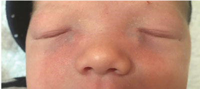
FIGURE 1

FIGURE 2
1. What is the most likely diagnosis?
- Congenital dacryocele (dacryocystocele)
- Encephalocele
- Haemangioma
- Orbital dermoid cyst
- Extra-nasal glioma
- Ethmoid mucocele
2. What initial investigations would you consider, if any?
- Ultrasound
- CT/MRI
- None
- Inflammatory markers
3. In this case, what would be the most appropriate initial management?
- ‘Watch and wait’
- Warm compress and massage
- Probing
- Topical antibiotics
- Systemic antibiotics
- Ophthalmology referral
Answers
1. The correct answer is (a)
Bilateral congenital dacryocele is the most likely explanation. This presents as a blue tinged tense cystic swelling located below the medial canthus of the eye.1 25% of cases are bilateral.2 It is caused by accumulation of mucus or amniotic fluid in the lacrimal sac due to obstruction of the nasolacrimal duct.1 The lower end of the duct is last to canalise, often opening after birth and sometimes not at all.
The remaining answers are possible differential diagnoses although unlikely to be bilateral. The most important to exclude is encephalocele – the herniation of intracranial contents through a skull defect. This is differentiated from dacryocele by location above the medial canthal tendon. The swelling is pulsatile and may be associated with displacement of the globe, more marked with crying.3
Haemangioma is a cause of tumours of the orbit and periorbital area. If deep in the preseptal area it may appear as a dark blue swelling. It may enlarge with crying or when the infant is held in a head-down position, but pulsation and bruit are absent.3 Haemangiomas are also generally less firm to the touch.
An orbital dermoid cyst is a firm, non-tender, well-circumscribed, mobile mass usually in the superior orbit, commonly the superotemporal area.3
Extra-nasal glioma is another rare cause of swelling around the eye that can present in infancy. It causes a firm red-blue mass that is non-pulsatile and does not enlarge with crying.4
Ethmoid mucocele occurs due to paranasal sinus obstruction. It is very rare and may present with proptosis.
2. The correct answer is (c)
The diagnosis here was made clinically with no further investigations. If there is diagnostic uncertainty, imaging may be helpful. Ultrasound might confirm a cystic mass; more detailed imaging may be obtained with CT or MRI.5 Investigation with blood tests for inflammatory markers is not well described. Occasionally nasal endoscopy is indicated if an associated nasal mucocele compromises the airway.
3. The correct answer is (a) and (b)
In the first instance it would be reasonable to ‘watch and wait’, with massage and warm compress to aid canalisation of the nasolacrimal duct. This leads to resolution in many cases.6
Further intervention may be required if there are complications. Early involvement of the Ophthalmology Department aids prompt assessment and management to prevent long-term morbidity.7 With infection (dacryocystitis), a combination of pharmacological and surgical intervention may be advocated. Topical antibiotics may be useful for conjunctivitis. If there is diffuse erythema and swelling of the overlying skin and eyelids, many paediatricians would start systemic antibiotics. If antibiotics do not help then an intervention becomes necessary to decompress the lacrimal sac.8 Probing and irrigation, or in some cases marsupialisation, may be helpful.9
Summary
A dacryocele is a cystic swelling located below the medial canthus of the eye that is caused by the obstruction of the nasolacrimal duct. The diagnosis of congenital dacryocele is usually made clinically but imaging with ultrasound, computerised tomography or magnetic resonance imaging may be useful in cases of diagnostic uncertainty. A ‘watch and wait’ approach is reasonable initial management in uncomplicated cases. Early involvement with ophthalmologists in complicated cases is important to prevent long-term morbidity.
Outcome
The patient was discussed with the Ophthalmology Department and effective massage technique was re-iterated with the parents. Chloramphenicol eye ointment was prescribed as a precaution. The condition resolved within two weeks with no complications.
Parental consent
Consent for publication was obtained from the patient’s family.
Contributions and acknowledgement
RS provided the idea for the article and identified the patient. CC and FW wrote the first draft. RS and KM reviewed the manuscript and made corrections. All authors reviewed the manuscript and agreed on the final version for submission.
With thanks to family of the infant for allowing us to share his story.
Or read this article in our
Tablet/iPad edition
