

A newborn with wrinkly skin: what is the diagnosis?
Connective tissue disorders present with widespread manifestations involving the skin, joints, ligaments, blood vessels and other organ systems and can be associated with significant disability and life-threatening complications. Important disorders in this group include cutis laxa, wrinkly skin syndrome and geroderma osteodysplasticum. This article reports on a newborn baby with geroderma osteodysplasticum to raise awareness of this rare condition and the challenges faced by affected children and their families.
Chikkanayakanahalli M. ManjunathaConsultant Neonatologist1
manjunatha.c@royalehayat.com
Ahmed Abourahma
Neonatal Registrar1
Samuel E. Ibhanesebhor
Consultant Neonatologist1 and Professor of Paediatrics and Neonatal Medicine2
Ashok Z. Mathews
Consultant Neonatologist1
1Neonatology and Newborn Nursery Department, Royale Hayat Hospital, Hawally, Kuwait
2Department of Paediatrics, Igbinedion University, Okada, Nigeria
The case
A full-term female baby was born at 39 weeks’ gestation by spontaneous vaginal delivery to a 25-year-old gravida 2, para 1 mother. She was in good condition at birth with Apgar scores of 9 at one and five minutes of age. The amniotic fluid was meconium stained but the baby did not require any resuscitation. The cord pH was 7.31 with a base deficit of -2.7. The baby’s birth weight and head circumference were appropriate for her gestational age. The birth weight was 3,060g (25th centile), head circumference was 34cm (50th centile) and length was 50cm (50th centile). Soon after delivery, the baby was noted to have abnormally loose and wrinkled skin.
The pregnancy was complicated with oligohydramnios but there was no history of diabetes or any other significant health issues of concern. It was a consanguineous marriage (second degree relatives). In view of consanguinity the mother, had non-invasive prenatal testing (NIPT), which was reported as low risk for aneuploidies and microdeletions.1 An antenatal anomaly scan was unremarkable except for hyper-extended neck. There was no history of any genetic or dermatological disorders in the family.
A detailed examination of the baby showed inelastic wrinkled skin all over the body (FIGURE 1), sagging cheeks, wrinkles radiating from the angle of the mouth and the chin, a receding mandible (FIGURE 2), bilateral talipes calcaneovalgus deformity, hyper-extended wrists with long fingers (FIGURE 3) and dysplastic nails in both feet. The left hip was dislocated and the right hip was dislocatable. The skin was inelastic, as evident by very slow return to normal after stretching.
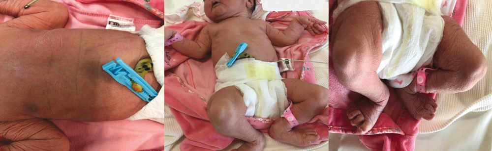
FIGURE 1 A detailed examination of the baby revealed inelastic wrinkled skin all over the body.
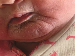
FIGURE 2 The face showed sagging cheeks, wrinkles radiating from the angle of the mouth and chin, and a receding mandible.
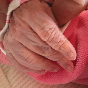
FIGURE 3 Hyper-extended wrists with long fingers.
The examination of the cardiovascular system showed normal heart sounds, no murmur and normal femoral pulses with a blood pressure of 65/34 (mean=44mmHg). The baby, who was not in respiratory distress, had a normal-shaped chest with equal air entry into both lung fields. Her abdomen was normal with no organomegaly and no umbilical or inguinal hernia. The spine was normal with no scoliosis.
Examination of the central nervous system revealed normal tone, normal deep tendon reflexes and anterior fontanel (fontanelle). There was a capillary haemangioma over the sacro-coccygeal region. The eyes were normally shaped with normal red reflexes. A newborn hearing screening by a brainstem auditory evoked response was normal.
The child’s parents were informed of the clinical findings and the need for further investigations and multidisciplinary input, including genetics and long-term follow up.
An echocardiogram on day 6 of life showed a small atrial septal defect (ASD) in addition to a patent foramen ovale (PFO), which resolved on subsequent scans. An electrocardiogram (ECG) showed normal sinus rhythm. Cranial and abdominal ultrasound scans were normal. Ultrasound scans of the hips on day 11 showed a femoral head outside the joint cavity on the right side and a subluxated left femoral head.
A genetic analysis of this baby at approximately three months of age identified a homozygous frameshift insertion mutation in the GORAB gene on chromosome 1q24, which is linked with geroderma osteodysplasticum.
At 7.5 months of age the child developed a fracture of the right tibia. She is now 10 months old with no dentition, she is unable to crawl and has constant tearing of both eyes. Her hip dislocation was managed with splints and she is awaiting surgery.
Discussion
There are more than 200 heritable disorders of connective tissue2 that are caused by genetic mutations affecting individual components such as collagen and elastin, or the enzymes regulating their metabolism. Collagen, a connective tissue protein, acts like glue giving strength and support to the body and elasticity for movement. Thus, the altered collagen affects the mechanical properties of the skin, joints, ligaments, blood vessels and other organs. Important disorders in this group include cutis laxa, Ehlers-Danlos syndromes (EDS), Marfan syndrome and pseudo-xanthoma elasticum. These disorders present with widespread mani-festations involving the skin as well as other systems, eg cardiovascular, skeletal and ophthalmological.3 Hence, all of these disorders can be associated with significant disability and life-threatening complications. Defining the disease phenotypes for these rare disorders is challenging because a phenotype that is thought to be a single entity, may in fact represent multiple conditions, each with a distinct, but overlapping presentation and clinical course.4
We suspected a connective tissue disorder in this baby because of her strikingly wrinkled skin appearance at birth involving the whole of the upper limbs, face and abdomen, along with the dislocated hip and joint hyper-extensibility. The differential diagnoses considered were congenital cutis laxa and EDS, due to the extensibility of the skin. Pseudo-xanthoma elasticum was eliminated because here the skin changes usually develop in early adulthood, although it can occur in childhood.
Infants with the classical-type EDS have hyper-extensible skin, which extends easily and snaps back after releasing it because of increased elasticity. Their skin feels soft and velvet-like; a feature that is lifelong. The skin texture has also been described as doughy.5 Hyper-extensibility of the skin is also a feature of cutis laxa, but here the skin is inelastic and resumes its normal shape slowly, as seen in this case. Cutis laxa may be congenital or acquired; in this article we will discuss the congenital form, which is relevant to this case report. To our knowledge, there are no accurate incidence data and there is no known racial or ethnic predilection.
Congenital cutis laxa
Congenital cutis laxa is a group of rare, inherited connective tissue disorders in which the skin becomes inelastic and hangs loosely in folds such that patients develop a prematurely aged appearance. Although any part of the body may be affected, the loose folds of skin are most prominent around the eyes, face, neck, shoulders and thighs with decreased elastic recoil on stretching, as in this case. Although the baby did not have hypotonia or flat feet as described in the literature, her right hip was dislocated and her left hip was unstable. Downward slanting palpebral fissures, a long philtrum, inguinal and umbilical hernia have been reported in cutis laxa, however, they were absent in this case. Skin fragility, easy bruising and poor wound healing are not associated with cutis laxa, unlike similar skin disorders. Some patients may develop diverticula of the small and large bowels and rectal prolapse. Many patients will develop pulmonary complications, such as emphysema, due to the loss of the elastin support network; these are the most common causes of death in these patients. Cardiovascular manifestations such as cardiomegaly, congestive heart failure, murmurs, cor pulmonale and aortic aneurysms may occur.
Geroderma osteodysplasticum
Geroderma osteodysplasticum is a rare autosomal recessive connective tissue disorder that, along with wrinkly skin syndrome, was traditionally included in the cutis laxa syndrome in view of its somewhat similar clinical features.6 Due to the many phenotypic similarities of these conditions, it was proposed that they represented the same disorder.7 Traditionally these conditions were differ-entiated based on radiological findings, clinical progression and differences in clinical maifestations.6 However, with advances in genetics these conditions can now be differentiated early at the molecular level by identification of mutated genes, as in this case report.
Consanguinity is reported frequently in families who have offspring diagnosed with wrinkly skin syndrome, cutis laxa with growth and developmental delay, and geroderma osteodysplasticum.6 Consanguinity increases the risk of autosomal recessive disorders; in this case, father and mother were second degree relatives (uncle and niece).1
The genetic analysis of this baby showed a homozygous frameshift insertion mutation in the GORAB gene.8 This gene encodes a member of the golgin family, a group of proteins localised to the Golgi apparatus. Mutations in the GORAB gene have been associated with geroderma osteodysplasticum, an autosomal recessive disorder characterised by:9
- changes in the skin suggesting precocious aging (wrinkled skin)
- generalised connective tissue weakness
- infantile-onset osteoporosis leading to frequent fractures (this child developed fracture at 7.5 months of age)
- underdeveloped cheekbones and jaw (malar and mandibular hypoplasia)
- a varying degree of growth deficiency and short stature.
Other features described in the literature include:9
- microcephaly
- droopy face
- relative prognathism (protruding jaw)
- deep-set eyes
- osteopenia
- progressive kyphoscoliosis, vertebral collapse, platyspondyly (in adults) and irregular endplates (in adults)
- bowed tibia, bowed femur
- camptodactyly, hyper-extensible fingers
- speech delay and mental retardation.
Many of these facial and other features were absent in this child.
Conclusion
This case shows the importance of meticulous clinical examination of all neo-nates to identify any anomalies or unusual appearances, considering differential diagnoses by correlating other clinical findings. Nevertheless, the patient discussed in this case report demonstrates the limitations of clinical examination in differentiating rare conditions with similar clinical features – here the importance of genetic evaluation is emphasised. Considering the complexity of genetic inheritance and possible implications for future pregnancies, it is important to counsel the parents sensitively. Babies with lifelong conditions should be referred to tertiary centres with adequate facilities for investigations and long-term multidisciplinary management.
Parental consent and acknowledgement
The authors are grateful for the support of the parents for consenting to write this case report and providing additional information, including progress reports.
Or read this article in our
Tablet/iPad edition
- Geroderma osteodysplasticum, cutis laxa and wrinkly skin syndrome are rare conditions that may be confused with Ehlers-Danlos syndromes.
- Defining the disease phenotype for these disorders is challenging because of overlapping presentations and clinical course.
- Careful clinical examination and genetic evaluation are important in differentiating rare conditions with similar clinical features.
