

Cleft palate and oral synechiae in a newborn infant
Cleft lip or palate associated with fibrous bands, also known as oral synechiae, is rare. This report describes a newborn infant with four fibrous bands in the oral cavity, a left-sided cleft lip and a cleft of the hard and soft palate. The fibrous bands made feeding difficult and raised concerns about the baby’s airway as they prevented normal positioning of the tongue.
Laura MorrisMBChB, MRCPCH, DTMH
ST5 Paediatrics
laura.morris18@nhs.net
Sanjeev Rath
MBBS, MD, FRCPCH
Consultant Neonatologist
Shri Babarao
MBBS, MD, DCH, FRCPCH, MSc, PGCME
Consultant Neonatologist
Neonatal Unit, Wirral Women and Children’s Hospital, Merseyside
The case report
It was first noted that the fetus might have a cleft lip on the 20-week anomaly scan of the 24-year-old primiparous woman. Several attempts at a full detailed anatomical scan were unsuccessful. Views obtained at 26 weeks confirmed a left-sided cleft lip; at that time involvement of the palate was felt to be unlikely based on the images that had been obtained. The parents received counselling from the
fetal medicine team and were offered amniocentesis because of the association between cleft lip/palate and chromosomal abnormalities. Amniocentesis was declined by the parents. A neonatal alert was generated with a plan of a senior member of the team attending the delivery and the baby staying with the mother unless there were other reasons to admit to the neonatal unit. The parents were made aware of feeding problems encountered in babies with cleft lip/palate.
The baby was born by normal vaginal delivery at 40 weeks’ gestation and did not need any resuscitation. The attending practitioner at delivery noted the presence of a left-sided cleft lip and fibrous bands extending from the lower alveolus to the roof of the mouth (FIGURE 1).
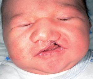
FIGURE 1 The left-sided cleft lip is clearly visible.
It was difficult to fully examine the mouth but the tongue was seen behind the bands and the movement of the tongue was restricted. A nasogastric tube was passed on the postnatal ward for overnight feeding. Following review by the cleft team in the morning the patient was noted to have a left-sided cleft lip and palate and four fibrous bands joining the lower alveolar mucosa to the roof of the mouth (FIGURE 2).
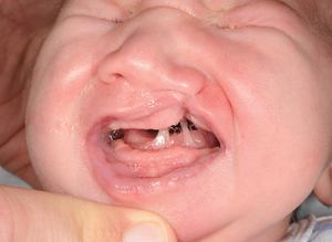
FIGURE 2 The left-sided cleft lip and fibrous bands. The tongue can be seen behind the fibrous bands.
The patient was transferred to the neonatal unit because of concerns that the baby’s tongue could obstruct the airway, and if the baby vomited it could cause aspiration due to the position of the tongue. The patient was kept nil-by-mouth and given intravenous fluids. Following discussion with the maxillofacial team the patient was urgently transferred to the regional tertiary surgical centre. Surgical intervention at a few hours of age divided the bands and confirmed the presence of hard and soft palate clefts. Immediately after surgery the baby was able to feed, and continued to establish feeding from a soft teat bottle with an assisted feeding technique. Since discharge the patient has been gaining weight adequately and remains under follow-up with the cleft team.
Review and discussion
Fibrous bands in the oral cavity are rare; there have been approximately 60 cases reported worldwide.1 If the bands are composed solely of soft tissue the condition is termed synechiae, and if there is bony involvement it is termed syngnathia.2 Some syndromes are associated with fibrous bands in the oral cavity, notably Van der Woude, Pierre Robin and popliteal ptrygium syndrome. Non-syndromic oral synechiae with an isolated cleft palate is even more rare; in 2002 Dalal found just five cases reported in the literature.3
Abnormalities occurring during embryological development have been incriminated in the pathogenesis of oral synechiae in a variety of different theories.4 It has been postulated that during normal oral development the tongue moves downward and forward in the seventh week to allow for fusion of the palatal shelf. Protrusion out of the oral cavity prevents fusion between the oral mucosae. The most widely accepted theory establishing the formation of oral synechiae is that they are the remnants of the buccopharyngeal membrane, as proposed by Mathis in 1962.5 The presence of a cleft palate in some children with synechiae has led to suggestions that these are a sequence: that the presence of an abnormal membrane prevented normal movement of the tongue in development and therefore prevented fusion of the palate.4
There are varying degrees of oral synechiae and Ogino et al6 describe five types:
- adhesion of the alveolar mucosa to one or both sides of the upper and lower jaw (alveolar synechiae)
- membranous adhesion on the hard palate and floor of the mouth, excluding the rear of the tongue (lateral synechiae)
- bands partially involving the hard palate and tongue
- bands in which the soft palate and tongue are widely involved
- bands involving a membranous adhesion between the hard palate and lower lip.1
In our index patient, management issues secondary to difficulty in feeding, concerns about airway compromise and risk of aspiration were considered appropriately because of the tongue being pushed backwards and associated with restricted movement. Other reports have highlighted difficulties with anaesthetising patients for surgery: some choose to perform it under local anaesthetic and sedation1,2,7 while others choose to use no anaesthesia at all.3 In our index case local anaesthesia was used to divide the bands.
Cleft palate is one of the most common congenital abnormalities, with approximately 1,200 babies born with a cleft each year in the UK.8 While a number of babies with oral synechiae also have a cleft palate, the number of babies with cleft palate who also have synechiae is very small. Rai et al found that of the 1,134 cases of cleft palate that they saw over a period of four years, there were only two with synechiae2 – an incidence of 0.18%.
Conclusion
The presence of fibrous bands in the oral cavity associated with cleft palate is very rare. This case highlights the importance in identifying the potential problems of difficulty in feeding, airway obstruction, increased risk of aspiration and difficulty in securing a definitive airway if the baby deteriorates. As a consequence, it should alert the medical team to mobilise the patient to the maxillofacial team for urgent surgical input.
It is such an infrequent finding that even tertiary centres rarely see babies with such a presentation, thus making it important to continue to raise awareness of early management.
Patient consent
The authors received written consent to publish this report from the patient’s parents.
Or read this article in our
Tablet/iPad edition
- In cases of fibrous bands the patient should be examined carefully for a cleft palate and associated syndromes.
- The baby is likely to need additional support with feeding and fluid management prior to division of fibrous bands.
- Fibrous bands can be associated with airway or breathing difficulties.
